Thursday, August 24, 2006

Haem- Bone Marrow

The technologist’s task in bone marrow aspiration procedures is to carry out on-site smearing of patient’s marrow. Usually there are 3 different types of smears done:

1) Direct aspirate/ wedge smears:
-Performed by placing a few marrow particles at one end of a clean slide and smearing with a spreader.
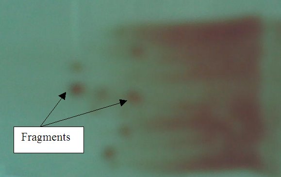
-Marrow particles (fragments) must be found mostly at the tail of the smear.
-After staining slides with MayGrunwald Giemsa stain, the fragments appear bluish in colour.
-When viewing under a microscope, a trail of cells can be seen following the fragments. Differential counts are performed in these cellular trails.
-Fragments are examined to determine the cellularity.

Wedge Smear

2) Squashed Preparation
-A few particles (fragments) are firstly placed on a slide. Another slide is then gently compressed on the first slide to spread and disperse the particles.
-The crushed fragments will be at the center of the smear.
-To see the cells of the fragments more clearly. (since the fragments are more spread out when it is squashed)

3) Trephine Imprint (from bone)
-Several (2-4 slides) touch or imprint preparations were made by gently touching the core plug (bone) and rolling it between 2 slides.
-This smear or preparation is especially useful if the aspirated marrow is diluted or a “dry tap” (i.e. no marrow particles or fragments can be extracted) is obtained.
-When ‘rubbing’ the core plug between slides, the cells on the surface of the core plug will be attached to the slide.

Other possible presence of cells to take note or when viewing marrow slides:
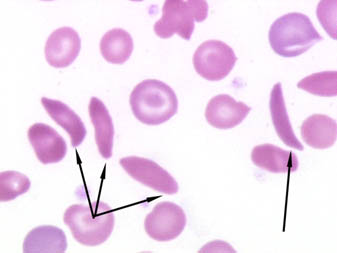
-Nucleated red blood cells (nRBCs)
-Megakaryocytes
-Mitotic cells (undergoing division)

Confirmatory tests in relation to possible diagnosis:
1) Iron stain (demonstrate presence of hemosiderin/storage iron in erythroblasts/sideroblasts or erythrocytes): useful in the classification of anemia associated with defective Hb synthesis. Can also be used to identify presence of ringed sideroblasts in the diagnosis of soderoblastic anemia and myelodysplasia.

2)Neutrophil Alkaline Phosphatase (NAP score): Used nto distinguish chronic granulocytic leukemia (CGL) from other myeloproliferative disorders and Polycythaemia Rubra Vera from secondary polycythaemia.

3)CSF test for blast cells: used to examine leukaemic cells in the CSF from a cytospin preparatrion. Essential in the diagnosis of meningeal leukaemia. Also used to monitor the adequacy and effectiveness of treatment.

4) Periodic Acid Schiff (PAS) reaction : stains a variety of carbohydrates including glycogen (found in haemopoietic cells). Used in differential diagnosis of acute leukaemia (e.g: Lymphoblasts are PAS positive.

Monday, July 24, 2006

Haem case study 2

Case: Thalassemia-related disease

Sample Collection: Whole blood via venipuncture
Samples Processing: Blood sample is collected in an EDTA tube (there must be at least 3mL of blood). Once received, a lab no. is given to the sample for identification purposes.

Preliminary laboratory tests: Alkaline Cellulose Acetate Electrophoresis (ACAE), Variant test.
The ACAE test is used to screen for Hemoglobin (Hb) variants. Hb have a net negative charge at pH 8.6 and move towards the positive electrode in an electrical field. The variation in the amino acid content of the different Hb causes variation of net charge of each Hb type. This hence determines their rate of mobility. ACAE makes use of cellulose acetate strips. When run at 350 volts for 25 minutes, the Hb bands will appear accordingly on the strips.
Procedure:
1) Blood samples are firstly washed with 0.9% NaCl and centrifuged. This step is done 2 times. The purpose is to get packed cells (pellet).
2) A fixed amount of washed packed cells is then pipetted into test tube containing hemolysate reagent.
3) The mixture is mixed and left to stand for 20 mins.
4) Meanwhile, prepare the cellulose acetate strips. It is labeled accordingly and placed in a carrying rack. The rack is then slowly lowered (to prevent formation of air bubbles) into a container containing supre-heme buffer and soaked for 5 mins.
5) Prepare sample wells. The patients’ hemolysate mixture (from step 3) and also the control (ESFA) is placed into each sample wells.
6) The applicator is primed by pressing gently the tips into sample wells 1-2 times. Check if the samples are well picked up by the applicator by applying it on filter paper.
7) It is then applied to the cellulose acetate strip.
8) The strip is then quickly placed in electro chamber (containing supre-heme buffer).
9) Electrophoresis the plate for 25 mins at 350 volts.
10) Staining: Ponceau S stain for 5 mins, 5% acetic acid for 2 mins, absolute methanol for 2 mins, cleaning solution for 5-10 mins.

Interpretation:
If there’s high amount (dark band) of Hb A2, this may indicate beta thalassemia as since there is lack of beta-chains, there’ll be excessive alpha chains causing high amounts of Hb A2. Confirmatory tests should follow.

An example of a gel strip.

The variant test is used to quantitate and detect HbA2 and HbF. It is intended for the separation and area percent determination of HbA2 and HbF, and also assists in the identification of abnormal Hb in whole blood using ion-exchange high performance liquid chromatography (HPLC).

Preliminary investigation: Haemoglobin levels, HbA2, HbF

Confirmatory tests:
1) Sickling test (can be used if the HbS band appears in ACAE test)
- Gives a rapid screening test for the Hb S status of a patient.
- Demonstrate the sickling phenomenon in test samples. Sickling phenomenon occurs with low oxygen tension. A small drop of blood is added to a reducing agent. It is then sealed between slide and cover glass, and incubated at 37 degree Celsius for sickling to occur. A negative and positive control is included together with the other samples. If there is no sickling in the positive control, this indicates false negative result hence test have to be repeated.


Arrows indicates sickle cells.

2) Acid elution test (Kleihauer)
- Used to determine the distribution of fetal Hb in red cells (confirmatory test when there’s an unusual Hb F band in the ACAE test.)
- Hb F resist acid elution while Hb A is denatured under these conditions. Thin blood films are exposed to low pH acid buffer, washed and then stained. Cells containing Hb F are deeply stained while cells containing Hb A gibe a ghost-like pale appearance.

3) Acid gel electrophoresis
- Enables separation and provides confirmation of Hb variants that co-migrated in alkaline Hb-electrophoresis. (e,g: to differentiate Hb C from Hb E when theres a dark band indicating Hb E in ACAE test).
- Hemolysate is applied to an acid buffered agarose gel. Electrophoresis then takes place. Following that, the Hb in the gel is immobilised in a fixative solution and the gel is dried to a film. To see the Hb pattern clearly, the gel is stained with a protein specific stain.

4) Hb H test
-The Hb-H test is performed to screen for Hb-H inclusion bodies. It can be used as a confirmatory test in the diagnosis of alpha-thalassemia.
-2 drops of brilliant cresyl blue stain is added to 100 microlitres of blood. After incubation period of 1 hr at 37 degree celcius, samples are left to cool before smears are made for microscopy viewing. Presence of Hb-H is indicated by “golf-like” cells.

Monday, July 03, 2006

Haem case study

Female, 57 years old
Given diagnosis: Acute lymphocytic leukaemia (ALL)

Routine FBC / Reference range
WBC --> 2.16 X 10³ cells/mL /4.5 – 11.0 X 10(power 9)
RBC --> 3.04 X 10⁶ cells/mL /4.2 – 5.4 X 10(power 12)
Hb --> 9.5 g/dL /11.5 – 16.0
HCT --> 27.2% /37% – 47%
MCV --> 89.7 fL /80 -96
MCH --> 31.3 pg /27-29
MCHC -->34.9 g/dL /31.5 – 34.5
RDW --> 15.6% /11.5% - 14.5%
PLT --> 21 X 103 cells/mL /150 – 450 X 10(power 9)

Routine WBC differential (100%) /Reference range
Neutrophil : 38 /40-75
Lymphocyte: 47 /15-41
Monocyte: 15 /2-10
Eosinophil : 0 /0-6

Morphology flags
Blasts : +
ATYP(atypical lymphocyte): +

Discussion:

Clinical presentation/ symptoms : Fatigue, weight loss, anemia, bleeding gums, breathlessness, enlarged lymph nodes, liver and/or spleen, fever and paleness.
Sample Collection: Whole blood via venipuncture
Samples Processing:
1)Blood sample is collected in an EDTA tube
2)A blood smear is made followed by staining. This is for microscopic viewing.
3)The blood sample is then placed on a special rack on the automated analyser to carry out full blood count (FBC)

Preliminary laboratory tests: FBC
Preliminary investigation: RBC, WBC, Hb, HCT, MCV, MCH, MCHC, RDW, PLT, differential count and morphology flags.
Expected results for laboratory tests:
1)Values of WBC, RBC, Hb, HCT and PLT are lower than reference range
2)Values of MCH and RDW are higher than reference range
3)Values of MCV and MCHC are normal.
4)There are also abnormal amounts of neutrophils, lymphocyte and monocyte.
5)There are presence of blasts (lymphoblasts) and atypical lymphocyte (ATYP)

Interpretation:
1) Values of WBC and PLT are low, indicating that there is failure of the bone marrow. In ALL, the WBC is usually high. In this case, the patient may have gotten leukaemia for quite some time hence the WBC may decrease due to earlier treatment.
2) The low amounts of RBC, Hb, HCT indicates anemia, which is one of the symptoms of leukaemia.
3) The presence of blasts highly indicates possibility of ALL.
4) An increased level of RDW reveals presence of anisocytosis due to bone marrow failure.
5) The abnormal amounts of WBC differential count confirm that there is abnormality of the WBC.

Confirmatory tests:
1) Cell markers test (flowcytometer) to confirm presence of lymphoid or myeloid.
2) Cytochemical stain: Used in the identification of the different subtypes of acute leukaemia.
3) Immunological markers (Antigens/ antibodies specific to ALL): A positive test indicates presence of ALL
4) Bone marrow aspiration / biopsy: An increased number of cells and lymphoblasts confirms presence of ALL.

Prognosis: There are treatments for leukaemia. This includes chemotherapy, steroids, radiation therapy and growth factors. The main goal of treatment is remission of the cancer. Administration of blood products (packed RBCs, platelets) may also be needed to treat the anemia and low platelet count.